
MBM Trainee Mikhail Kandel recently published an article titled “Epi-illumination gradient light interference microscopy for imaging opaque structures” in Nature Communications.
The article, published in October 2019, details how Kandel et al. built a new kind of microscope for imaging miniature brain machinery. Other contributors in the MBM Program include trainee Kathryn Sullivan and faculty Hyunjoon Kong, Martha Gillette, and Gabriel Popescu.
The work represents a collaborative effort between a few groups within the MBM Program, addressing challenges in imaging samples provided by Drs. Gillette and Kong.
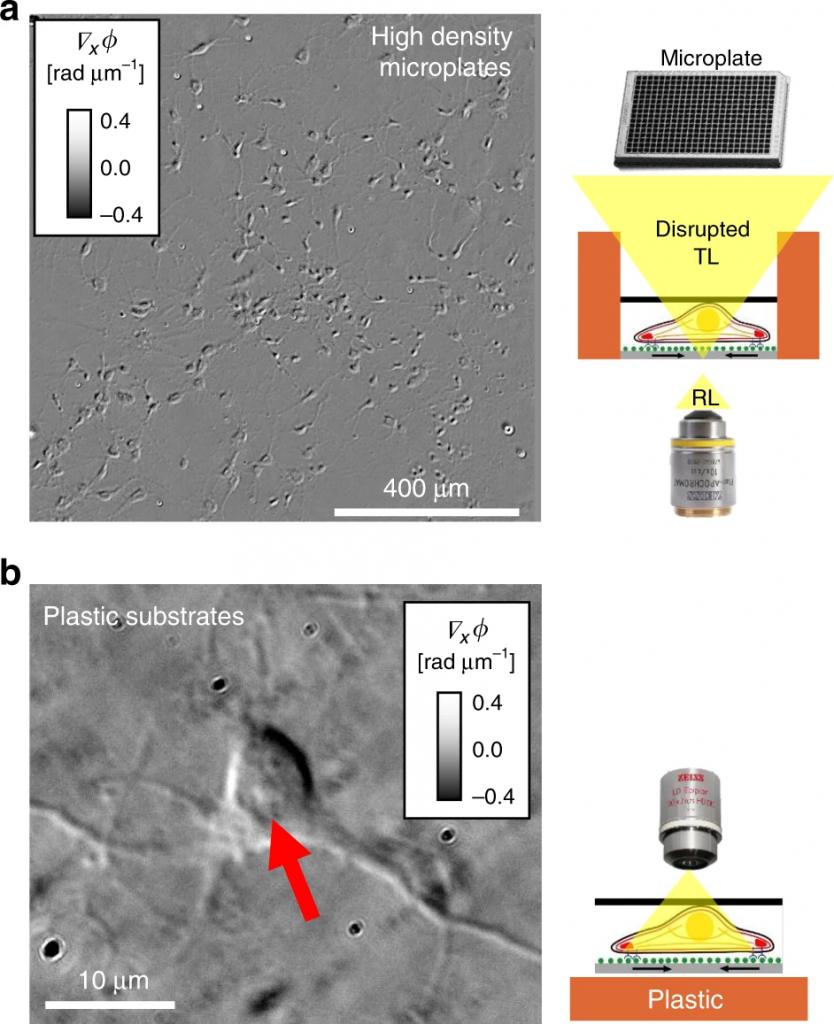
“I think that this work enables high throughput, label-free quantitative phase imaging for personalized medicine applications,” explains trainee Mikhail. “By imaging in reflection, we can record cells growing on microtiter plates (Figure 3 above) where each miniature brain would receive a different dosage of some chemical.
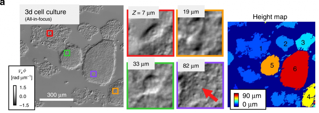
Figure 5a (below) illustrates these kinds of images.
“This was very much an imaging paper, and among the rare times people measured the back scattering signal which potentially has higher resolution. To perform 3D reconstruction we came up with a new technique to measure the impulse response of the microscope by using beads, which is a small step forward in one of the grand challenges in our field (3D object reconstruction),” Mikhail added.